Overview
Craniopharyngioma is a rare type of noncancerous brain tumor.
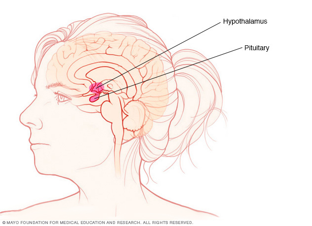
Craniopharyngioma begins as a growth of cells near the brain's pituitary gland. The pituitary gland makes hormones that control many body functions. As a craniopharyngioma slowly grows, it can affect the pituitary gland and other nearby structures in the brain.
Craniopharyngioma can happen at any age, but it occurs most often in children and older adults. Symptoms include changes in vision over time, fatigue, headaches and urinating more often. Children with craniopharyngioma may grow slowly and may be smaller than expected.

Diagnosis
Diagnosing a craniopharyngioma usually starts with a medical history review and a discussion of symptoms. Tests used to diagnose a craniopharyngioma include:
- Neurological exam. During this exam, a health care professional tests vision, hearing, balance, coordination, reflexes, and growth and development. This can help show which part of the brain might be affected by the tumor.
- Blood tests. Blood tests may reveal changes in hormone levels that show a tumor is affecting the pituitary gland.
- Imaging tests. Imaging tests capture pictures of the brain. The pictures can show the size and location of the tumor. Imaging tests include X-rays, CTs and MRIs. In certain situations, other tests might be needed.
Treatment
Craniopharyngioma treatment often starts with surgery. When possible, surgeons remove all of the tumor. Sometimes the craniopharyngioma can't be removed completely. Surgeons may remove as much of the craniopharyngioma as is safe.
Radiation therapy may be used after surgery to treat any tumor cells that remain. Using surgery and radiation together often provides good tumor control. This approach also helps lower the risk of complications after surgery.
Other treatments, such as chemotherapy and targeted therapy, might be options in certain situations.
Surgery
Most people with craniopharyngioma have surgery to remove all or most of the tumor. What type of surgery you have depends on the location and size of the tumor.
- Open craniopharyngioma surgery. Also called a craniotomy, this operation involves opening the skull to access the tumor.
- Minimally invasive craniopharyngioma surgery. Also called a transsphenoidal procedure, this operation uses special surgical tools inserted through the nose. The tools go through a natural passage to get to the tumor. This approach minimizes the effect on the brain.
When possible, surgeons remove the entire tumor. However, there are often many delicate and important structures nearby. This means surgeons sometimes can't remove the entire tumor. To ensure a good quality of life after the operation, surgeons remove as much of the tumor as possible. Other treatments may be used after surgery.
Surgeons do the operation in a way that avoids hurting nearby structures during the operation. Surgeons take care to reduce the risk of vision problems. They work to minimize damage to the hypothalamus. The hypothalamus helps with many functions, including controlling appetite and alertness.
Surgery is sometimes used to relieve a fluid backup on the brain, called hydrocephalus. A tube can be placed to drain the fluid. Often the tube is temporary. Sometimes a permanent tube is needed. A permanent tube, called a shunt, can drain the brain fluid to the abdomen.
Radiation therapy
Radiation therapy uses powerful energy beams to control tumor cells. The energy can come from X-rays, protons or other sources.
Radiation therapy is often used after surgery to treat any tumor cells that are left.
Types of radiation therapy for craniopharyngioma include:
-
External beam radiation therapy. During external beam radiation therapy, you lie on a table while a machine moves around you. The machine directs radiation to precise points on your body.
Specialized external beam radiation technology allows your health care team to carefully shape and aim the radiation beam. This helps to deliver treatment to the tumor cells and reduces the chances of hurting healthy tissue. These technologies include proton beam therapy and intensity-modulated radiation therapy, also called IMRT.
- Stereotactic radiosurgery. Stereotactic radiotherapy is an intense form of radiation treatment. It aims many beams of radiation from many angles at the tumor. Stereotactic radiosurgery treatment is typically completed in one or a few treatments.
- Brachytherapy. Brachytherapy involves placing radioactive material directly into the tumor where it can radiate the tumor from the inside.
Chemotherapy
Chemotherapy uses medicines to kill tumor cells. Chemotherapy can be injected directly into the tumor so that the treatment reaches the target cells. This makes it less likely to damage nearby healthy tissue.
Treatment for papillary craniopharyngioma
Targeted therapy medicines might be a treatment option for one type of craniopharyngioma called papillary craniopharyngioma. This type isn't common. In adults, about one out of every three craniopharyngiomas is the papillary type.
Targeted therapy uses medicines that attack specific chemicals in the tumor cells. By blocking these chemicals, targeted treatments can cause tumor cells to die.
Nearly all papillary craniopharyngioma cells contain a change in their DNA called the BRAF gene. Targeted therapy aimed at this change may be a treatment option. Lab testing can show whether your craniopharyngioma contains papillary cells. Tests also can tell whether those cells have the BRAF gene change.
Clinical trials
Clinical trials are studies of new treatments. These studies provide a chance to try the latest treatments. The risk of side effects might not be known. Ask your health care team if you might be able to be in a clinical trial.
© 1998-2025 Mayo Foundation for Medical Education and Research (MFMER). All rights reserved. Terms of Use


