UM Health-Sparrow Comprehensive Breast Center
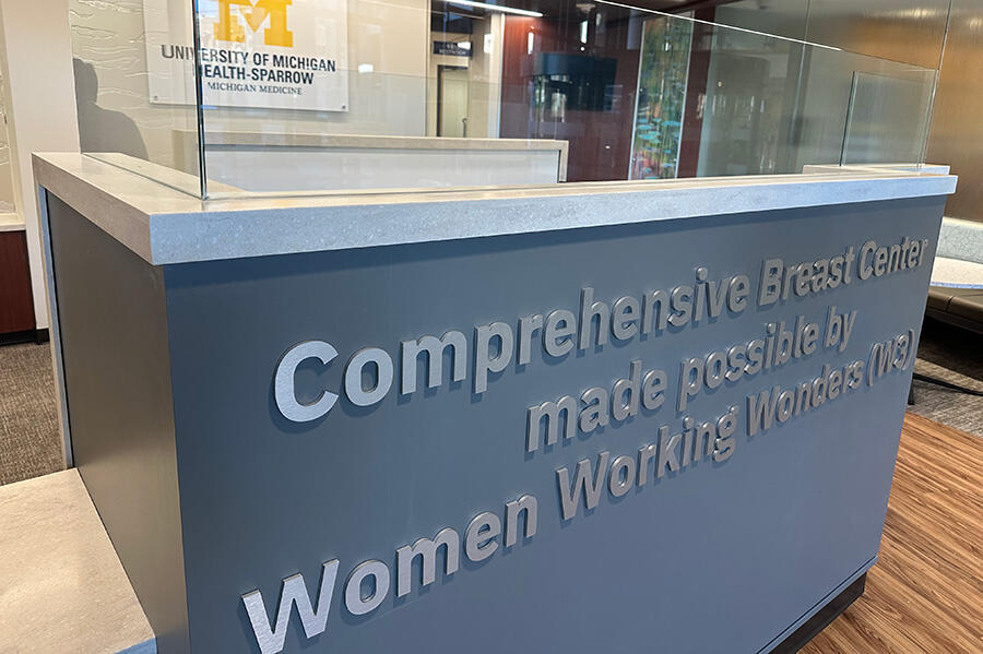
University of Michigan Health-Sparrow has opened an expanded Comprehensive Breast Center.
The 9,000-square-foot center, located on the first floor of the Sparrow Professional Building at 1215 E. Michigan Ave., Lansing, MI, unites breast health services in one accessible location. It aims to improve the patient's experience by eliminating the need to travel to multiple locations for screening, diagnostics and treatment coordination. The facility features innovative technology, including ultrasound, stereotactic biopsy and mammography machines with 3D technology, which is particularly beneficial for patients with dense breast tissue. This updated center also reflects the power of philanthropy and community partnership, with support from Women Working Wonders.
We support the recommendation of the American College of Radiology and Society of Breast Imaging, which advocates for annual clinical breast exams with your primary care physician and annual mammograms starting at age 40. These are the best tools in detecting breast cancer in its earliest and most treatable stages. UM Health-Sparrow Comprehensive Breast Center is a proud accredited member of the National Accreditation Program For Breast Centers (NAPBC).
Schedule an Appointment
Our experienced healthcare professionals have specialized training and are dedicated to your breast health; we are ready to answer your questions, perform your annual mammogram, or guide you through a more serious diagnosis. Plus, we offer a comfortable space and privacy during your exam.
To schedule your mammogram, call 1-800-698-6329.
UM Health-Sparrow Eaton Breast Care Center
Eaton also offers comprehensive breast health services. In addition to screening and diagnostic 3D digital mammograms, they offer other services, including bone density to genetic testing.
For more information or to schedule a mammogram at Eaton, call 517-541-5948.
Clinical Services
Screening Mammogram
A routine, annual mammogram, reviewed by a board-certified radiologist, and compared to any previous breast images to watch for areas of concern.
Diagnostic Mammogram
This occurs when a concern appears on your screening mammogram, or if you have a particular sign or symptom that warrants a more in-depth investigation. This exam will usually be focused on a certain area of your breast.
Breast Tomosynthesis (3D Mammogram)
Using digital mammography, multiple images are taken to create a 3D picture of the entire breast. This tool is an advanced screening that provides greater detail and clarity to detect 30–40% more breast cancers in women with dense breasts.
Breast Ultrasound
Waves that are used to create an image of the breast tissue. This can be used in combination with a screening or a diagnostic mammogram.
Magnetic Resonance Imaging (MRI)
This tool provides screening for certain women who are at a higher risk for cancer through a personal history, a strong family history, or for women with breast implants.
Breast Intervention Services
Our experienced and caring staff and comprehensive services offer hope and encouragement. Every patient and every finding is unique. While women travel different paths through our breast intervention services, the goal is early identification, intervention and treatment.
- Cyst Aspiration Fluid: collected from a cyst in your breast for analysis.
- Ductography: ordered if you are having an abnormal discharge from your breast to determine the possible origin.
- Breast Biopsy Small: samples of your breast tissue are collected from the area of concern for pathology analysis.
- Fine Needle Aspiration Biopsy: a small needle is used to withdraw cells from one or more suspicious areas in your breast.
- Needle/Wire Localization: a very small, fine wire is inserted to mark the exact location of an area of concern and serve as a guide to the surgeon. This is done when you arrive at the Breast Center, just prior to your surgery.
- Sentinel Lymph Node Tracing: a low-dose radioactive tracer is injected just under your skin near the site of concern and traced to the first lymph node where it appears, called the sentinel node. The surgeon will remove this node to test for cancer. This procedure determines if a cancer has spread and how far.
Preventative Testing
Screening Mammogram
A routine, annual mammogram, reviewed by a board-certified radiologist, and compared to any previous breast images to watch for areas of concern.
Diagnostic Mammogram
This occurs when a concern appears on your screening mammogram, or if you have a particular sign or symptom that warrants a more in-depth investigation. This exam will usually be focused on a certain area of your breast.
Breast Tomosynthesis (3D Mammogram)
Using digital mammography, multiple images are taken to create a 3D picture of the entire breast. This tool is an advanced screening that provides greater detail and clarity to detect 30–40% more breast cancers in women with dense breasts.
Breast Ultrasound
Waves that are used to create an image of the breast tissue. This can be used in combination with a screening or a diagnostic mammogram.
Magnetic Resonance Imaging (MRI)
This tool provides screening for certain women who are at a higher risk for cancer through a personal history, a strong family history, or for women with breast implants.
Breast Intervention Services
Our experienced and caring staff and comprehensive services offer hope and encouragement. Every patient and every finding is unique. While women travel different paths through our breast intervention services, the goal is early identification, intervention and treatment.
- Cyst Aspiration Fluid: collected from a cyst in your breast for analysis.
- Ductography: ordered if you are having an abnormal discharge from your breast to determine the possible origin.
- Breast Biopsy Small: samples of your breast tissue are collected from the area of concern for pathology analysis.
- Fine Needle Aspiration Biopsy: a small needle is used to withdraw cells from one or more suspicious areas in your breast.
- Needle/Wire Localization: a very small, fine wire is inserted to mark the exact location of an area of concern and serve as a guide to the surgeon. This is done when you arrive at the Breast Center, just prior to your surgery.
- Sentinel Lymph Node Tracing: a low-dose radioactive tracer is injected just under your skin near the site of concern and traced to the first lymph node where it appears, called the sentinel node. The surgeon will remove this node to test for cancer. This procedure determines if a cancer has spread and how far.
Locations
Make a Donation
If you would like to make a gift to support breast health services at the Comprehensive Breast Center at UM Health-Sparrow, please visit UofMHealthSparrow.org/Give or select the button below. Thank you for your support.


